BioImage Analysis
The DCSR offers image analysis support for the FBM community. Whether you're looking to extract quantitative information, automate a manual task, fix a script, or discuss a new project, you're at the right place! Antony Carrard, bioimage analyst and machine learning expert, will be happy to help you.
What we can work on together
From a quick question to a full pipeline build from scratch, if it involves extracting information from images, we can talk about it. Here is what I can help you with:
- Image acquisition & setup — camera and lighting selection, acquisition protocol, color calibration
- Pre-processing — denoising, stitching, channel alignment, flatfield correction
- Segmentation & detection — from classical thresholding to deep learning models (SAM, Cellpose, StarDist…)
- Cell & object identification — detection, counting, classification by morphology or intensity
- Colocalization — spatial co-occurrence analysis, intensity correlation, custom distribution metrics
- Quantification — intensity profiles, shape descriptors, color and texture analysis
- Tracking & behavioral analysis — object tracking across frames, movement and behavior metrics
- Large-scale pipelines — automation, batch processing, HPC when needed
- Results & visualization — overlays, plots, summary tables, exportable metrics
Common tools & software
Depending on your needs and existing workflow, we can also work with established open-source bioimage analysis tools:
- Fiji / ImageJ — the standard for general image processing and macro automation
- CellProfiler — automated quantification pipelines, no coding required
- QuPath — whole-slide and multichannel image analysis
- Ilastik — interactive machine learning for segmentation and classification
- Napari — Python-based viewer for multidimensional images
Projects
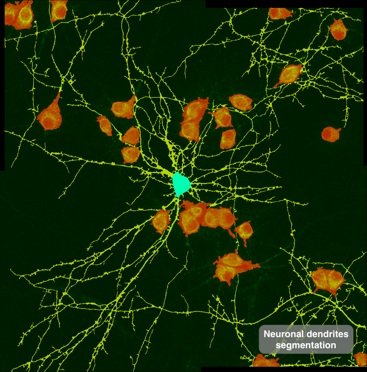
Automatic dendritic segmentation for intensity extraction — Marianna Pompili, Bagni Lab, DNF
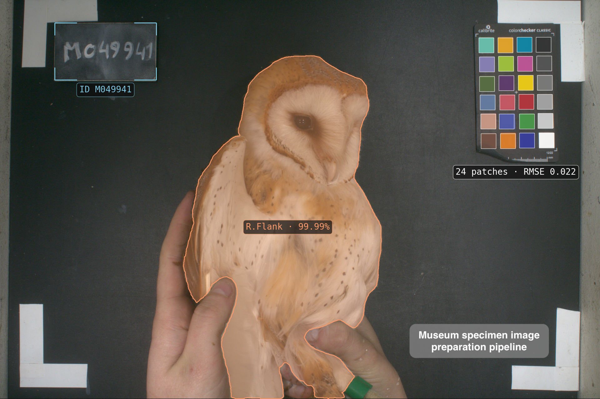
Owl specimen ID extraction and image color correction pipeline — Tristan Cumer, Goudet Group, DEE
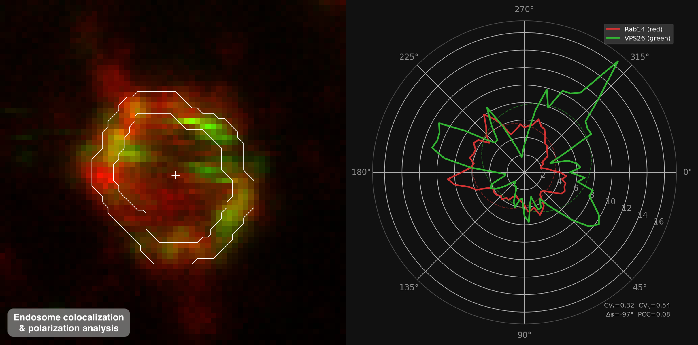
Automatic endosome contour identification for colocalization and polarization analysis — Sarah Hornfeck, Widmann Lab, DSB
Contact
Feel free to reach out at research-computing-fbm@unil.ch — briefly describe your project or question and we'll get back to you shortly.